This work was one of many conservation and remounting projects undertaken in 2014 and 2015 as part of the Grant Museum of Zoology’s
‘Bone Idols’ program of work (click on the link for more information about the project).
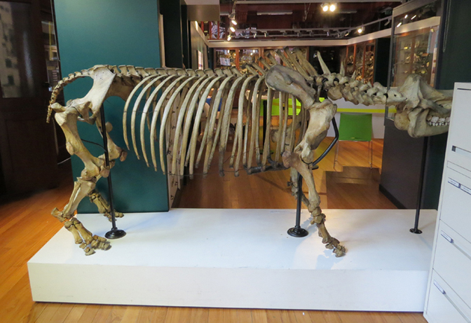
This articulated skeleton of an Indian one-horned rhinoceros was acquired by the museum over 100 years ago, but was not mounted particularly well at the time. The shape of the vertebral column was wrong, the skull was higher than the pelvis rather than vice versa, the two upright supports did not connect with the vertebral rod in a secure fashion and the weight of the skull and mandible pulled the front upright off vertical. Also, the skull did not sit on the front metal rod very well and was secured in place by an unsightly combination of a hook, screw and wire. Therefore the point of remounting the specimen was to: change the spinal column from a straight horizontal line to a more natural and anatomically correct curve; lower the neck and skull relative to the pelvis; have all four feet actually touching the plinth; have the head of each femur articulate with the acetabulum of the pelvis; and to improve the way the skull was held in place so that it did not pull the upright supports off vertical and was under less stress. The skull was not to be in a ‘grazing position’ nor to ‘be leading with the horn’.

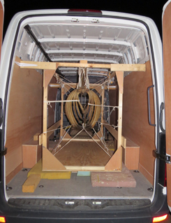 All the work was undertaken by natural history conservator Nigel Larkin, so the skeleton had to be partially dismantled and transported from central London
to his conservation workshop in the West Midlands. Taking the specimen apart and preparing it to undergo a long road journey safely was
quite a project in itself (see images to left and right), some of which was described by UCL staff here:
Dismantling Reg the Rhino in Ten Easy Steps including a time lapse video.
All the work was undertaken by natural history conservator Nigel Larkin, so the skeleton had to be partially dismantled and transported from central London
to his conservation workshop in the West Midlands. Taking the specimen apart and preparing it to undergo a long road journey safely was
quite a project in itself (see images to left and right), some of which was described by UCL staff here:
Dismantling Reg the Rhino in Ten Easy Steps including a time lapse video.
The whole skeleton was dusty and dirty, particularly the horizontal surfaces where dust and dirt had settled. In some places there was chalk or paint on the surface of the bone, and spots of white paint. A portion of the premaxilla had fallen off in the past and needed re-attaching, as did the head of the left femur.
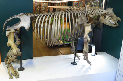

Above: The condition of the specimen before work started. Above left, the skeleton with the neck portion
incorrectly higher than the pelvis. Right, the front supporting pole no longer in a vertical position.
Cleaning The whole skeleton required cleaning as it was covered with dust, dirt and some paint and chalk (see images below).
The dust and some dirt was removed with a very soft brush used next to the hose of a vacuum cleaner to remove the debris.
Then all the bones were cleaned with a gentle conservation detergent, an alcohol ethoxylate (Synperonic A7) in distilled water.
It was applied to small areas at a time with a soft brush, then wiped away immediately with a lint-free paper towel.
Then the area was cleaned again with clean water (with no detergent) and immediately dried again with a paper towel.


Above far left: The neural spines and the transverse processes on the right of these vertebrae have been cleaned,
highlighting how dirty and dusty they still are on the left side of the vertebrae.
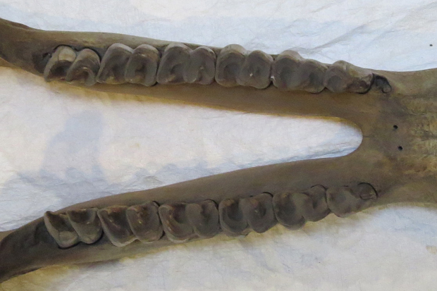
Above left: The teeth and bone of the mandible before cleaning. Above right, the same teeth and bone after cleaning. Above far right:
Paint on the underside of the mandible.
It is amazing what you can find when cleaning. Not only were remains of plants
found in the cavities and cracks of the teeth from the rhino's last meal (these were photographed and left in place
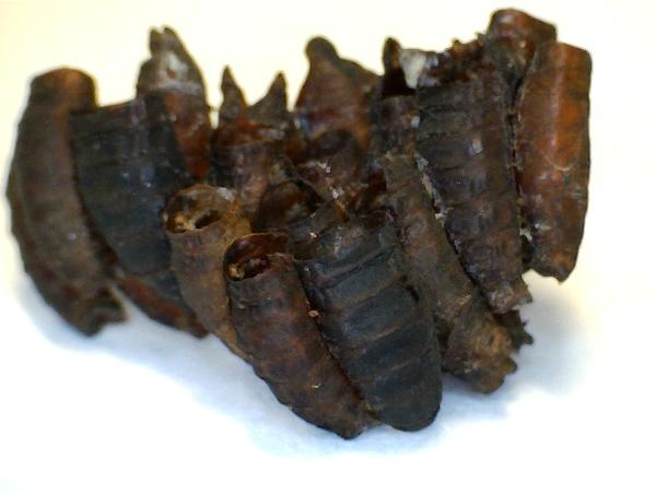
for future study) but some clusters of
insect coccoons were found in the nasal cavities. These were identified as 'rhino bot fly' – a rare parasite of live rhinos. See the Grant Museum's page
'The Return of the Rhino: Conserving our biggest skeleton'.
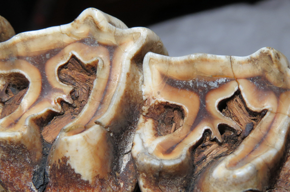
Above left: Some remains of vegetation still stuck within the teeth of the rhino.
Above right: some of the botfly larvae casts from the nasal cavity (photo copyright UCL Grant Museum of Zoology).
 Repairs The sternum had been modelled at some point in the past using some kind of putty or resin mixed
with fibres that may be hair. This has cracked extensively over the years and some small pieces had fallen off.
All the modelled material was consolidated (with Paraloid B72 in acetone) to strengthen it and to give a good surface
for the repairs to adhere to. Plaster of Paris was used to fill the worst cracks and model-in missing pieces.
This was cleaned with a small amount of dilute citric acid in water, then the plaster was painted with artists’ acrylic paints.
The head of the left femur was also rejoined to the shaft of the bone from which it had become separated and broken pieces were
rejoined to the premaxilla of the skull, all with the conservation adhesive Paraloid B72.
Repairs The sternum had been modelled at some point in the past using some kind of putty or resin mixed
with fibres that may be hair. This has cracked extensively over the years and some small pieces had fallen off.
All the modelled material was consolidated (with Paraloid B72 in acetone) to strengthen it and to give a good surface
for the repairs to adhere to. Plaster of Paris was used to fill the worst cracks and model-in missing pieces.
This was cleaned with a small amount of dilute citric acid in water, then the plaster was painted with artists’ acrylic paints.
The head of the left femur was also rejoined to the shaft of the bone from which it had become separated and broken pieces were
rejoined to the premaxilla of the skull, all with the conservation adhesive Paraloid B72.