Report on the cleaning, repair and conservation of Ichthyosaurus crassimanus prior to its redisplay in York Museum in March 2011
 This rare complete skeleton is the Type Specimen of Ichthyosaurus crassimanus, from an alum shale quarry
north of Whitby, donated to the museum by the Rev D. R. Roundell in 1857. It is nearly 30 foot long
and was originally displayed vertically, mounted on a wall secured with iron and copper bars from behind
and embedded in mortar and plaster. It was removed from the wall in 1991. During the process, evidence of extensive dry rot and woodworm was found within the mount material, and some pyrite decay within the specimen.
This rare complete skeleton is the Type Specimen of Ichthyosaurus crassimanus, from an alum shale quarry
north of Whitby, donated to the museum by the Rev D. R. Roundell in 1857. It is nearly 30 foot long
and was originally displayed vertically, mounted on a wall secured with iron and copper bars from behind
and embedded in mortar and plaster. It was removed from the wall in 1991. During the process, evidence of extensive dry rot and woodworm was found within the mount material, and some pyrite decay within the specimen.
Condition of the specimen
The postcranial material consisted of several dozen pieces, ranging in size from blocks weighing in excess of 50 kgs down to small sealed bags containing broken fragments of bone a centimetre or so across.
The specimen has many fine to medium-sized cracks that opened post-excavation. Some apparently opened up when the specimen was on display before. Building work in the museum in the late 1980s utilised percussion drills and it is thought some of these cracks may relate to the vibrations caused by this work. Four pairs of marker disks were attached either side of cracks in the abdomen in 1989 with polyester resin whilst it was still on display when there was concern about the cracks widening. These disks enable the movement of the specimen either side of the cracks to be measured, so they have been left in place to be of further use.
For more details about what we can do for you, or for a quote, please contact:
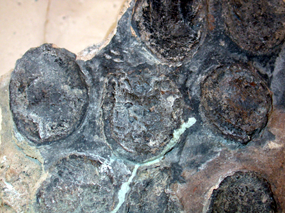
 Some cracks may have opened up since and although the specimen is not currently considered to be at risk from pyrite decay, in the 1970s the specimen seems to have been treated with morpholine for pyrite decay and whilst removing the specimen from the wall in 1991 it was found that the phalanges of one of the pectoral limbs (presumably the right pectoral limb, see below) had been affected quite badly by pyrite decay, making them very friable. The only area of pyrite decay found during the 2010/2011 conservation project was within the ribs of block C.

By-products of pyrite decay on block C on the left, and paint splatters on ribs on the right.
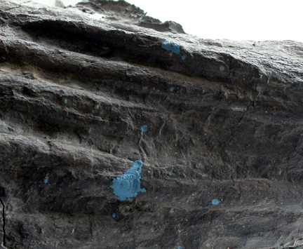
All the pieces still had a variety of mortar, plaster, wax and/or green epoxy resin (Araldite) attached to their sides and rears, from where gaps between the bones had been filled, or where pieces were ‘adhered’ together using these materials. Also, as the specimen was set vertically into a wall there was an excess of mortar, plaster and/or resin applied cosmetically to the edges. All this ‘foreign’ material had to be removed, so that the pieces were reduced to just rock and fossilised bone and clean enough that a good join or repair could be made between pieces. Large amounts of excess paint also had to be removed – blue and green paint from the edges where the surrounding area had been painted, as well as splatters of paint all over the skeleton from when the gallery had been redecorated over the years. Also, tissue paper had been consolidated to the specimen with polyvinyl alcohol before removing it from the wall in 1991 to help keep broken pieces together. This had to be removed along with 20 years’ worth of dust, dirt and fluff etc from when the specimen had been kept on open racking in the museum stores, partly covered in bubblewrap.
Various metal bars used for mounting the specimen to the wall were still attached to the rear of some blocks, inserted deep within them. These could not be pulled out so were sawn off flush with the rear of the specimen. The skeleton was prepared in the 1950s with the best techniques available at the time. This meant that there were areas of the specimen that benefitted greatly from further light preparation using small percussion tools and the airabrasive unit to enhance details.
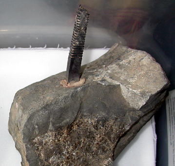

On the left, one of the copper spikes used to attach the specimen to the wall.
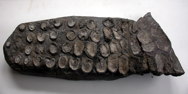
In the middle, phalanges of the right pectoral limb before preparation and cleaning - and afterwards on the right.
Materials used
All materials used in this conservation project are removable and/or reversible. The only adhesive used on this specimen during the conservation process was Paraloid B72 (a reliable and reversible museum-quality methacrylate copolymer), and the consolidant was the same, mixed with acetone (about 5% PB72, 95% acetone). Small gaps were filled with a mixture made from Paraloid consolidant (25% in acetone) and glass beads 44 microns in diameter (see Larkin & Makridou, 1999). This adhesive, consolidant and gap filler are all reversible with the application of acetone. The skeleton was rebuilt in manageable sections, with no section made to be heavier than two people can comfortably move together.
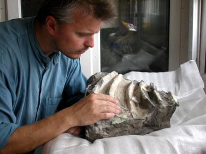
Cleaning and Preparation
Cleaning was undertaken with compressed air, gentle vacuuming, brushing with soft brushes, and with an airbrasive unit utilising sodium bicarbonate and a variety of small pneumatic pens and chisels. Every single piece was cleaned this way, removing large amounts of mortar, plaster, resin and wax to leave clean pieces consisting only of rock and fossilised bone. This means the specimen is much less adulterated with materials, and more details can be appreciated. Also, better adhesion was achieved when rejoining pieces. In addition, some areas of the specimen were lightly prepared in the process, resulting in giving much greater definition to some of the pieces so that the bones can be more greatly differentiated from their host matrix, facilitating a greater appreciation of the anatomy. This was particularly true of the phalanges of the limbs.
Examples:


Block U before, during and after cleaning.

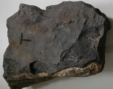


The rear of block T before and after cleaning on the left, and two pieces halfway through cleaning on the right.


A section of the tail showing the tissue to be removed, then the paint and mortar that needed removing.
The labels that had been attached either side of the cracks were all on the upper surface and would be visible to the public. Therefore they all had to be moved to the internal sides of the breaks to be much less visible. They were retained for future reference, in case the specimen is moved or studied at a later date.


On the left: Epoxy resin and mortar that had been used to fill an old gap. On the right: a label moved from the upper surface to the side surface (the white mark above the label shows where it was before).
Not all the epoxy resin filler could be removed. Sometimes it was the only clue as to how two blocks were to join together. In some cases, remove g the filler could not be done without damaging the specimen as the resin was often stronger than the rock and fossilised bone. Where the resin was left in place for these reasons, it was painted out.


Epoxy resin in the specimen on the left, and painted out on the right.

Removing tissue from a specimen, and using the airabrasive on one of the biggest pieces in a glovebox.
Restoration of the right pectoral limb
Three of the limbs had been broken in to several medium-sized pieces and consisted of fossilised bone and host matrix.
However, the distal half of the right pectoral limb was a mass of broken mortar and many
loose bones. The mortar in which they had been set had deteriorated badly and was completely falling apart.
Bones were matched up with holes in the mortar, or in the area where the mortar was missing they were arranged
in their correct positions according to the figure in the manuscript provided.
The decision was made to keep as much of the original mortar as necessary to make sure the bones stayed in their
original and correct positions, rather than rebuilding the limb from scratch and possibly lose some context.
However, the middle section did have to be rebuilt as the host mortar or original matrix was completely missing. This was possibly the result of pyrite decay observed in 1991 during demounting. Some of the phalanges were quite weak and friable.
After all the pieces has been cleaned, the mortar as well as the bones was consolidated to give it some mechanical
integrity. Then the pieces of mortar were adhered together and as many of the bones attached as possible.
Other bones were placed in their right positions and resin was built up around them.
The whole limb was backed with fibre glass cloth and resin to provide strength to the limb.
The original mortar extended far beyond the bones to make a very rectangular shape with straight edges that was completely different to the other three limbs. Therefore excess mortar was trimmed off with an angle grinder, taking care not to create too much vibration within the paddle.


Top left: Phalanges of right pectoral paddle in badly crumbling mortar, top right shows how the central portion has no matrix or mortar around the phalanges (including those on the bench in the background above the main paddle). Bottom left shows the rebuilt paddle before trimming and painting and bottom right shows the finished limb.
The left pelvic limb had a section of badly deteriorated mortar running through the centre. This had to
be rebuilt in a similar way, using resin with glass fibre cloth.


Left pelvic limb before cleaning, conservation and rebuilding (left) and after (right).
Mounting & installing
It was decided that no external mount was required for the specimen as it was to lay in a ‘sand tray’ in the display case. To get the various elements of the specimen in to position relative to one another, a combination of wooden blocks and polypropylene sand bags filled with dry sand were used as required to get the blocks to the right height and orientation. Cracks between some pieces were filled with sand to the required height. More sand was poured around the specimen and arranged accordingly to hide the supports under the specimen.
The skull, which required little in the way of conservation, was cleaned with soft brushes and a vacuum cleaner and then treated with a single application of Paraloid consolidant at 5% in acetone to bring out its colour and therefore make it similar in appearance to the rest of the consolidated skeleton.


The skull before consolidation on the left, and on the right after consolidation.
Installing the specimen in the gallery:


References
Larkin, N. and Makridou, E. (1999) Comparing gap-fillers used in conserving sub-fossil material. The Geological Curator 7 (2), 81-90.
Melmore, S. 1930. A description of the Type Specimen of Ichthyosaurus crassimanus, Blake (Owen MS). Annals and Magazine of Natural History, Ser.10, Vol. Vi, p615.
enquiries@natural-history-conservation.com
We are members of the United Kingdom Institute for Conservation of Historic and Artistic Works